| 
             Laboratory of Tumor Cell Biology. National Cancer Institute. NIH. 
              Bethesda. Maryland. USA  
            It is a particular honor for me o give the first Frederick Stohlman 
              memorial lecture because of my personal respect and friendship with 
              Fred. His courage. honesty. and stimulation of good science in the 
              hematological field will be remembered by all who knew him. It is 
              also very fitting to have these lectures in Wilsede where special 
              friends were made at the first meetings and where association and 
              friendship with Fred Stohlman became much closer for many of us. 
             
             
              Introduction  
            There have been enormous advances in the therapy of lymphocytic 
              malignancies as exemplified by the treatment and apparent cures 
              of some childhood acute lymphoblastic leukemias (see D. Pinkel elsewhere 
              in this book). The myelogenous leukemias remain much more difficult 
              to treat. and for this reason and because they also provide an interesting 
              hematopoietic system for the study of differentiation our interests 
              for the past several years has focused on the origin and pathogenesis 
              of this disease group. The origin of the myelogenous leukemias appears 
              to involve proliferation of astern cell with various degrees of 
              commitment to differentiation and arrests in differentiation at 
              various cellular levels. These diseases appear to be monoclonal 
              (see Fialkow and also Rowley elsewhere in this book) when they are 
              clinically manifest. However. it is not known if the initiation 
              of the disease is multiclonal or monoclonal. Their appearance as 
              monoclonal when presented to clinicians might be because of a select 
              growth advantage of one clone as often seen in tissue culture. We 
              do not yet know the mechanism(s) initiating the abnormality of growth 
              characteristic of these diseases. but certain factors have clearly 
              been shown in select cases to induce or play some role in leukemic 
              development. Studies in mice. chickens. and humans implicate genetic 
              factors which may be involved at multiple levels. Thus. some congenital 
              diseases with chromosomal abnormalities and certain families have 
              shown an unusual incidence of leukemia (see report by R. Miller 
              elsewhere in this book). One family of unusual interest has recently 
              been described by F. Gunz and his colleagues in Australia. Thirteen 
              members of the family developed some form of myeloproliferative 
              disease. clearly implicating genetic factor(s). Yet. some members 
              developed the disease within a relatively short time of each other 
              even though the age of each varied [17]. This certainly suggests 
              an environmental factor may have been operative as well as genetic 
              factor(s). There was no known unusual exposure of any member of 
              the family to chemicals or radiation. What about environmental factors? 
              Radiation can induce leukemias in animals" and it has been clearly 
              associated with myelogenous leukemia in humans under unusual circumstances. 
              Benzene also has been associated with leukemia in very rare instances. 
              and some other chemicals with such rarity that their leukemogenic 
              potential in man is undefined. Epidemiological studies indicate 
              that leukemia is not increased in people living at higher altitudes 
              with greater exposure to radiation" nor is leukemia generally higher 
              in industrial areas than in rural regions [24]. These observations 
              together with the facts that I" the incidence of childhood leukemia 
              has apparently stayed about the same since industrialization and 
              in fact has declined in recent years, 2" the incidence peaks in 
              young children. 3" the incidence is greater in whites than in black 
              people. and 4. association with chemicals and radiation is very 
              rare (see R. Miller elsewhere in this book) all suggest to me that 
              the leukemias may be predominantly biological diseases and that 
              all biological factors must be thoroughly explored. 
             
              Retroviruses and Retrovirus Related Information: Reasons for 
              Intensive Exploration of Human Tissues  
            We have felt that a search for retrovirus (RNA tumor virus" oncornavirus) 
              information or related information in human cells was mandated by 
              several considerations. I. As discussed above epidemiological considerations 
              are a stimulus for consideration of biological factors. Although 
              these ,same broad studies also do not lend strong support to a virus 
              causation of the disease in a conventional manner, I think they 
              are quite consistent with a role for viral information if one considers: 
              a) long latency" b) a second or third factor in addition to appropriate 
              viral information may be a requisite. c) retroviruses can be vertically 
              transmitted either in the germ line as an endogenous cellular element 
              or by congenital infection (see R. Weiss elsewhere in this book). 
              These factors would obscure epidemiological approaches. 2. Several 
              retroviruses can produce leukemia in a variety of animals in laboratory 
              experiments. 3. Retroviruses can sometimes transform cells and not 
              be seen again as discrete virus particles in in vitro experiments 
              (see P. Duesberg elsewhere in this book). Moreover" some data suggests 
              this may also be true with some naturally occurring leukemias of 
              animals. For instance. in a significant number of cats with leukemia 
              (perhaps approaching 50%), feline leukemia virus (FeLV) has not 
              been found (see 0. Jarrett elsewhere in this book). In many of these 
              cats antibodies to FeLV has also not been identified, and recent 
              data from our laboratory in collaboration with W. Hardy and M. Essex 
              indicate that FeLV proviral nucleic acid sequences may also not 
              be readily detectable (Koshy, Wong-Staal" Gallo, Hardy, and Essex" 
              in preparation). Yet there is evidence that FeLV may still cause 
              the disease in these so called "virus negative" cats. This is based 
              on some epidemiological results (M. Essex, personal communication) 
              and on the finding of feline oncornavirus membrane antigen (FOCMA) 
              in the leukemic cells. This protein is believed to he specifically 
              coded for or induced by FeL V and by feline sarcoma virus (FeSV) 
              (see M. Essex elsewhere in this book). 4. Bone marrow transplants 
              studies have indicated that in some exceptional cases normal hone 
              marrow donor cells may be transformed to leukemic cells when given 
              to a leukemic individual [43]. 5. We have recently been able to 
              show that some of the primate type-C retroviruses can transform 
              human Blymphocytes and may also interfere with differentiation of 
              myelopoietic cells, 6, The most compelling reason to think of retroviruses 
              in the etiology of leukemias ofhumans is the results of animal models. 
              It appears now that in every instance where we know the cause of 
              naturally occurring leukemia in a sizable fraction of a leukemic 
              animal population it involves a retrovirus. This now includes chickens 
              (see R. Weiss and 0. Jarrett, A. Burney elsewhere in this book), 
              some wild type mice [16], cows (see 0. Jarrett in this book), cats 
              (see Jarrett and also Essex) and gibbon apes (see next section). 
              7. Since retroviruses can recomhine with cellular genes and since 
              some of them can affect cell differentiation (see later section 
              of this report and also reports by M, Dexter, by N.Teich, by M, 
              Moore, and by T. Graffin this book), it is possible and perhaps 
              likely that sometimes these viruses contain cell derived genes involved 
              with growth and/or differentiation. If so then it should be important 
              to use retroviruses as probes in human leukemia to see if such genetic 
              information is altered during leukemogenesis whether or not the 
              disease is due to a virus. For this we would choose a primate retrovirus. 
             
             
              Primate Type-C Retroviruses  
             
             There were no isolates of any primate retrovirus before this decade. 
              Now there are many, and they are from diverse species. I will focus 
              on two groups since they were the earliest isolates and the only 
              ones which have been shown to have pathological effects on cells. 
              we have been particularly interested in the members of the infectious 
              type-C virus group isolated once from a woolly monkey and called 
              simian sarcoma virus (SiSV), simian sarcoma associated virus (SiSA 
              V) complex and some isolated several times f'rom gibbon apes and 
              collectively called gibbon ape leukemia virus (GaLV). SiSV (SiSAV) 
              and the various GaLV are very closey related viruses, and the evidence 
              suggests their ancestral origin Was probably a rodent virus which 
              entered these primates by interspecies infection [27,47]. It is 
              of particular interest that the virus entered two primates which 
              are only distantly related. We have especially focused our attention 
              on this virus group because the gibbon is the species closest to 
              man for which a retrovirus has been isolated, because it is the 
              species closest to man for which we have an animal model of leukemia 
              and notably one which we know something of the etiology, and because 
              we think viruses like this have been in humans. It  


              appears sufficiently clear that this virus entered gibbons directly 
              or via an unknown vector from a rodent, and it is certain that the 
              virus is transmitted by infection among gibbons in captivity. The 
              immediate vector to the one woolly monkey is unknown. A summary 
              of these viruses is given in Table I. Shortly after the isolation 
              of viruses of the SiSV-GaLV group [9,21,22, 39.42,46], baboon endogenous 
              type-C viruses (BaEV) were isolated from different tissues and different 
              species of baboons [20,40]. These viruses exhibit all the characteristics 
              of endogenous viruses, i.e., transmitted in the germ line of baboon 
              cells, and had no demonstrable effect in vivo or in vitro on mammalian 
              cells. Apparently BaEVentered an ancestor of domestic cats several 
              millions of years ago, infecting the germ line, and now maintained 
              as an endogenous virus of cats known as RD114 (reviewed by Todaro 
              in reference [41 ]). There is also evidence from several laboratories 
              that a related virus is sometimes identified in humans (see below). 
             
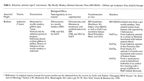
            Table 2. Endogenous Type-C Virus or Baboons (BaEV)a 


              a References to original reports (except tor recent result,) can 
              be obtained from either the text or the review by Gallo. R.C. and 
              Todaro. G.J. Oncogenic RNA Virus In: Seminars in Oncology Yarbro. 
              .JW. Bornstlein, RS, Mastrangelo. M.J (eds). pp 81-95 New York Grune 
              & Stratton. Inc. 1976  
             
              Selected Results with Human Cells  
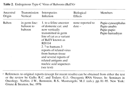
            Numerous reports suggest that some fresh uncultured human cells 
              contain intracytoplasmic virus like particles (discussed in references 
              [10] and [37]), but since these are not isolated as infectious virus 
              and have not been shown to exhibit biological activity it is not 
              certain thay they represent defective or abortive type-C viruses 
              or are cellular artifacts. However, the presence of high molecular 
              weight RNA with some sequence homology to RNA from some viruses 
              and, in some samples of human leukemic cells, a DNA polymerase with 
              properties resembling reverse transcriptase (RT), associated with 
              these "particles", emphasize their viral-Iike properties. In some 
              cases these polymerases were shown to be immunologically specifically 
              related to RT from SiSV-GaLV group (reviewed in references [10,11] 
              and [12]), but in most cases immunological relatedness to primate 
              viruses was not found and extensive comparisons (by immunological 
              tests) to other animal viruses was usually not performed because 
              of limited amount of enzyme. These results were a stimulus to consider 
              the possibility that humans may harbor viruses related to this group. 
              Our objective was to purify sufficient enzyme to enable us to make 
              peptide maps of the RT and compare this to RT from these viruses. 
              This has not been achieved, and there has been little progress in 
              our laboratory with this problem since the initial reports. On the 
              other hand Chandra and colleagues [1,2] have reported the purification 
              of this enzyme (related to SiSV-GaL V RT) from the spleen of a child 
              with a preleukemic disease, myelofibrosis, which eventually turned 
              into AML. Similar enzymes have been found in orbital chloromas from 
              leukemic people living near Ankara, a disease appearing as a cluster 
              (see R. Miller and also P. Chandra elsewhere in this book). On rare 
              occasions whole type-C virus has been reported isolated from human 
              cells. These reports, reviewed in references [II] and [13], have 
              come from our laboratory (HL23V), from Panem and Kirsten (HEL-12), 
              from Nooter and Bentvelzen and their colleagues (SKA-21), from Gabelman 
              and Waxman, and recently from H. Kaplan and colleagues. In each 
              case the viruses are related to the SiSV-GaL V group, and at least 
              in three cases (HL23V, SKA-21, and HEL-12), a second virus related 
              to BaEV has been detected. This peculiar combination remains unexplained. 
              Because of their similarity to existing primate viruses it is possible 
              that these isolates are all laboratory contaminants. Regarding HL23V 
              we offer the following results against contamination: I. Reproducible 
              isolation from separate clinical specimens: 2. previous evidence 
              reported in references [30] and [7] for RT related to RT of SiSV-GaLV 
              in the uncultured blood cells; 3. previous evidence for cytoplasmic 
              RNA sequences related to SiSV and BaEV in the fresh uncultured blood 
              cells of this patient [33]; 4. previous evidence for DNA proviral 
              sequences related to BaEV [33,48], although SiSV proviral sequences 
              could not be detected [33,48]. We did not subsequently find evidence 
              for a humoral antibody response to either the SiSV or BaEV component 
              of HL23V in the sera of this patient, but recently we have obtained 
              confirmation of the presence of the BaEV provirus in the DNA of 
              the uncultured blood cells from this patient. As described elsewhere 
              in detail in this book [49], we have used the technique of DNA digestion 
              with a restriction endonuclease followed by separation of DNA fragments 
              by agarose gel electrophoresis, transfer of the DNA fragments to 
              nitrocellulose filters by the Southern blotting technique, hybridization 
              of I125-labeled 35S viral RNA to this DNA, and examination for virus 
              specific bands after development over X-ray films. This approach 
              allows for more sensitive molec ular hybridization because much 
              of the irrelevant DNA is excluded (the labeled viral RNA probe is 
              in excess) and for qualitative assessment because the positive bands 
              can be visualized and compared to bands of virus infected cells. 
              As shown elsewhere in this book [49] DNA from fresh uncultured blood 
              cells from patient HL-23 and from another AML patient labeled HL-49 
              contain several clearly visible bands after digestion with the endonuclease 
              HIND III which hybridize to l12.5-35S RNA from BaEV (M7). For control 
              purposes DNA from normal human leukocytes and from human cells (A204) 
              deliberately infected by BaEV are also shown. No viral specific 
              fragments with BaEV as a probe were found in the normal cell DNA. 
              The only band seen is from ribosomal DNA which was detected with 
              labeled rRNA which was used as a control because sometimes rRNA 
              can contaminate viral RNA. Multiple viral specific bands are, or 
              course, found in the positive control, BaEV infected A204 cells. 
              It is interesting that some of the proviral bands found in the DNA 
              from the leukemic cells of patients HL-23 and HL-49 are not found 
              in the BaEV infected A204 cell positive control. This suggests that 
              the integration sites may be different or that the viral fragments 
              detected in HL-23 and HL-49 are related but not identical to the 
              M7 strain of BaEV. Despite the evidence for some cytoplasmic RNA 
              sequences related to SiSV (SiSA V) in HL23 fresh blood cells [33] 
              no novel proviral bands of SiSV (SiSA V) were found. M. Reitz in 
              our laboratory had previously shown that DNA from the kidney and 
              spleen of patient HL-23 contained more SiSV (SiSA V) hybridizable 
              sequences than did DNA from normal cells or DNA from the leukemic 
              blood cells of patient HL-23. It is possible that only a small percent 
              of the cells contained the SiSV (SiSA V) proviral sequences, and 
              failure to detect them is due to insufficient sensitivity. Such 
              cells may have been concentrated in the kidney and spleen. Unfortunately, 
              DNA or tissue from these organs is no longer available so they cannot 
              be examined by the restriction endonuclease blotting technique. 
              In summary, we have evidence now that the BaEV component of HL23V 
              was present in the primary uncultured blood cells of patient HL-23, 
              but inconclusive data for the SiSV (SiSA V) component. The important 
              question regarding these or similar viruses is to ask whether they 
              could be important causes of human leukemia. In an effort to obtain 
              a preliminary answer to this question we surveyed DNA from many 
              human tissues for the presence of integrated novel viral sequences 
              by the standard techniq ues of molecular hybridization. We used 
              molecular probes (both labeled RNA and labeled cDNA) from many animal 
              viruses, including SiSV and BaEV. We did not find significant differences 
              between normal and leukemic cells with probes from the majority 
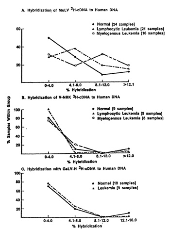
              of viruses, and the level of hybridization were insignificant. An 
              example of this is the negative data obtained with a rat type-C 
              endogenous virus and with GaLV illustrated in Fig. 1 (parts B and 
              C). The data is a summary of results of many samples presented as 
              a distribution frequency, i.e., it represents the percent of human 
              DNA samples (ordinate) which hybridize to a certain maximum extent 
              (abscissa). Note that the vast majority of cases (normal and leukemic 
              hybridize very little to rat virus nucleic acid probes and the distribution 


              Fig.l. Survey of human DNA from normal blood and from 
              people with various types of leukemia for murine leukemia virus 
              (MuLV) (IRauscher strain) related sequences (panel A). rat endogenous 
              virus (V-NRK) related sequences (panel B). and gibbon ape leukemia 
              virus (GaLV) related sequences (panel C) The probes are 3H-cDNA 
              prepared from endogenous reverse transcriptase reactions of the 
              respective viruses Data are given as the frequency and extent of 
              hybridization found in the various DNA samples As shown all samples 
              hybridized less than 8% ofcDNA probes from VNRK and GaLV. and there 
              are no differences between normal and leukemic samples These hybridization 
              resulls are not significantly above background levels Therefoore. 
              within the limits of sensitivity of the assay such sequences could 
              not be detected in humans In contrast some human DNA samples did 
              hybridize some of the MuLV probe to levels which appear to be significant 
              These results do not discriminate between MuLV sequences themselves 
              or sequences related to MuLV This survey was carried out by N Miller 
              and M. Reitz in our group.  
             
              of samples is virtually identical with DNA from normal and leukemic 
              cells. In contrast, using BaEV probes we find a small number of 
              leukemic DNA samples which hybridize significantly more of the probe 
              (data not shown). We have not conducted an extensive survey for 
              BaEV related sequences using the more sensitive approach of restriction 
              endonuclease Southern blotting, but we have applied it to a few 
              very select cases and verified the higher hybridization obtained 
              in the molecular hybridization survey. These results were mentioned 
              above and presented with AML  


              Fig.2. Survey or human DNA from normal blood and from 
              people with various types or leukemia for Simian sarcoma virus (woolly 
              monkey) (SiSV) related sequences In the upper panel (A) the probe 
              used was 3H-cDNA prepared from endogenous reverse transcriptase 
              reactions In the lower panel ( B) the probe was 1 125 labeled 70S 
              RNA. In both cases the vast majority or DNA ,samples from normal 
              tissue, hybridized less than 8% and all but a few less than 16% 
              In contrast a significant percent or leukemic samples hybridized 
              more than 12% and several more than 16% Again. these results do 
              not discriminate between SiSV sequences and SiSV related sequences 
              This survey was carried out by M. Reitz and N Miller In our group 
                
             
              patients HL-49 and HL-23 elsewhere in this book [49]. If the survey 
              results are a true indication of the presence of these sequences 
              then we can safely rule out BaEV or a closely related virus as commonly 
              associated with human leukemia and therefore as a common cause of 
              human leukemia. However. i t ma y be important to keep in mind that 
              the results could be underestimations. e.g.. if only fragments of 
              provirus arc present and/or only a small number of cells in a population 
              contain viral sequences. the sequences might easily be undetected. 
              but possibly suficient for leukemic transformation. We also surveyed 
              DNA purified from many human tissues for SiSV related sequences, 
              and as illustrated in Fig. 2 several leukemic DNA samples hybridized 
              more nucleic acid probes of SiSV (grown in marmoset cells) than 
              did DNA from normal tissues. This is more clearly evident when one 
              compares the distribution of hybridization ofviral probes to various 
              human DNA samples between rat virus (Fig. I) and SiSV (Fig. 2). 
              However, one DNA sample from a normal tissue (one of six human placentas 
              tested) also showed unusually high hybridization to SiSV probes, 
              so clearly higher hybridization is not unique to certain leukemic 
              cells. To verify these higher hybridization results and to obtain 
              greater sensitivity, some of these samples were also analyzed by 
              restriction endonuclease digestion-Southern blotting, and hybridization 
              of the specific fragments to SiSV 35S RNA. As described elsewhere 
              in this book [49], the positive results with the one placenta derived 
              from a normal person (termed NP3) obtained in the standard molecular 
              hybridization survey was confirmed as was one leukemic sample. In 
              these cases it is difficult to escape the conclusion of' infection 
              by an SiSV related virus. The presence in placenta is particularly 
              interesting since the finding suggests the possibility of congenital 
              virus transmission, a mechanism apparently involved in natural virus 
              transmission seen in chickens [44]. cats [19], cattle [26], and 
              gibbon apes [23]. The data are also consistent with the isolation 
              of an SiSV related virus from a human embryo by Panem and Kirsten 
              [32]. As noted earlier SiSV and GaLV are not endogenous to primates 
              but present in them only after infection. Although their recent 
              history (? vectors) is unknown their ancestral origin is believed 
              to be rodents. An unexpected observation. that came out of the search 
              of human DNA for inserted viral sequences related to SiSV and GaLV 
              was the finding of an SiSV related sequence in all hum{1n DNA. This 
              was detected after digestion of the DNA with BAM I, with HIND III, 
              and with Xpa restriction endonucleases (see paper by F. Wong-Staal 
              et al. elsewhere in this book). This sequence was specific to this 
              virus group since no endogenous viral sequence (i.e. sequences in 
              DNA from normal uninfected tissues from various cell samples) were 
              detected with probes from other viruses. We found this sequence 
              using either SiSV (SiSA V) or SiSA V, and the results were obtained 
              with viruses grown in non-human cells. Therefore, the results cannot 
              be due to a trivial explanation such as presence of human cellular 
              sequences with the viral probe. Moreover. it appears that this is 
              not an evolutionarily conserved sequence since it was not found 
              in the DNA we have tested from other normal uninfected primates. 
              We, therefore, tentatively interpret these results to indicate that 
              this group of viruses has been in the human population or an ancestor 
              of humans.  
             
              The Possibility of an Immune Response to Type-C Viruses in Humans 
             
            One of the most important questions relevant to type-C viruses 
              and humans i" whether a serological response against them is detectable. 
              Results presented previously [25,38] and elsewhere in this book 
              by R. Kurth and also by H. Snyder and by N. Hogg and their colleagues 
              deal with this complex and as vet unsettled issue. We recently explored 
              a different approach. Stimulated by the fIndings of I. Witz and 
              his colleagues [45], P. Jacquemin, C. Saxinger, and I examined human 
              blood cells for surface immunoglobulins. Both IgG and IgM were found, 
              and appear to be chiefly associated with non-lymphocytic cells, 
              a finding in agreement with Metzgar et al. [29] and Cotropia et 
              al. [5]. We discovered that among the IgG were some which react 
              with high specificity and at low concentrations with purified reversc 
              transcriptases (RT) from select mammalian leukemia typeC viruses. 
              In AML we find the IgG generally reacts with RT from SiSV, and to 
              our surprise the reaction can be specific enough to distinguish 
              this RT from the RT of the other members of the SiSV-GaLV group. 
              In normal people (bone marrow) we find about 20% positive for IgG 
              chiefly reactive with RT from one of the GaLV isolates, namely GaLVsI. 
              In CML we find the remission and chronic phase patients to be like 
              normal (negative or reactive with RT from GaL VsF), while in almost 
              every CML in blast crisis the IgG is chiefly reactive with RT from 
              FeLV and rodent type-C viruses [18,36]. Unlike the sporadic detection 
              of apparent inserted viral nucleic acid sequences the finding of 
              IgG with specific RT enzymatic neutralizing activity is common. 
              Because of this and because we have not as yet dcvcloped an assay 
              independent of RT activity neutralization,l it is premature to conclude 
              that this IgG represents an immune response to a viral protein. 
              It is possible that the IgG is directed against determinants of 
              a protein coincidentally very close to the amino acid sequences 
              of the catalytic sites of certain RTs. The answer to this question 
              should come from the isolation and characterization of the putative 
              cell surface antigen.  
             
              Growth and Differentiation of Human Leukemic Myeloid Cells and 
              Effects of Primate Type-C Viruses  
            We have reported that in the presence of conditioned media (CM) 
              from certain human embryo cell strains, human myelogenous leukemic 
              cells can often be induced to grow in liquid suspension tissue culture 
              [8,14]. In many instances the cells terminally differentiate [14,15]. 
              Sometimes they retain marker chromosomes, and this observation combined 
              with the fact that this CM generally does not induce growth of normal 
              myeloid cells led us to conclude that the maturation arrest of many 
              human myelogenous leukemias is not always irreversible, in agreement 
              with L, Sachs [35] and M. Moore [31] and their colleagues. The differences 
              in response of normal myeloid cells and leukemic cells also suggested 
              to us that membrane receptors for regulation may be modified in 
              leukemia. An interesting by product of this research was the establishment 
              of the first human cell line with distinct myeloid characteristics 
              [3]. This line, called HL-60, was shown to consist mostly of promyelocytes 
              [31] which can be terminally differentiated when DMSO or other compounds 
              active in the Friend murine erythroleukemia system are added to 
              the culture [3].  
             
              Effect of Primate Type-C Viruses on Normal Fresh Human Blood and 
              Bone Marrow Cells  
            To our knowledge there are surprisingly no reported studies of 
              the effects of primate type-C viruses on fresh human blood cells. 
              We have recently completed a preliminary study of the effects of 
              various type-C viruses, including the primate viruses described 
              here on human blood and bone marrow cells. We find that SiSV (SiSV), 
              SiSA V alone, GaLV, HL23V (SiSV component), but not BaEV induce 
              growth (Fig. 3) of B-Iymphocytes (Fig. 4). In about one third of 
              cases studied, the cells become immortalized, and despite apparent 
              normal karyotype, they are tumorigenic in nude mice (Fig. 5). These 
              cells are Ig and EBNA positive and do not have myeloid or T-cell 
              characteristics [28]. The observation is different from that of 
              "spontaneous" transformation of EBV positive human B-Iymphocytes 
              because the induction of growth is faster, the frequency of establishment 
              of cell lines in our hands much greater, and the cells are tumorigenic 
              despite apparent normal chromosomal makeup. The addition of BaEV 
              or FeLV (despite some virus replication in a few cases) did not 
              produce this effect. Finally, recent results indicate that the same 
              phenomenon can be induced with EBV negative cord blood but at a 
              lower frequency. We conclude that by indirect or direct means and 
              by mechanisms not yet understood, some primate type-C viruses can 
              be involved in the in vitro transformation of human B-Iyrnphocytes. 


              Working Hypothesis  
            A currently interpretable and simple model for leukemogenesis is 
              that the leukemogenic event whatever the cause, leads to auto-production 
              of a growth promoting molecule which prevent binding of normal regulatory 
              molecules. This model is similar to one proposed by Todaro and colleagues 
              in the genesis of sarcomas [6]. The alternative is that receptors 
              for regulators are themselves modified. These ideas should become 
              testable in the very near future.  


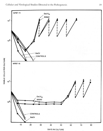
            Fig.3. induction of growth of B-Iymphocytes from 
            normal human blood by primate type-C retroviruses (Gal" VH and SiSA 
            V). Human blood leukocytes were cultured with or without the Hall's 
            Island strain of GaLV or with the simian sarcoma associated virus 
            (helper virus) (SiSAV). In some cases (e.g.. sample NPB 7-i5 shown 
            in the figure) induction of growth was rapid In other cases (e.g., 
            sample NPB 7-16 shown in the figure lower panel) growth induction 
            occurred after 50 days Spontaneous growth (no virus) occurred in only 
            1 to 2% of samples but in more than 40% of cells treated with viru5 
            if a co-cultivation method was used (P Markham. F. Rucetti. Z. Salahuddin. 
            R Gallagher, and R. Gallo. in press).  


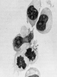
            Fig. 4. Morphology of „immortalized cell line induced from 
            normal human blood after infection by Ganot IVH and EBV positive ( 
            Magnification 1000 x ) 


              Fig,5. B-Cell lymphoma of nude mouse induced by 
              injection of mouse with cells induced to grow by GaLVH Cells are 
              human as determined by karyotype analysis. (From P Markham, F Ruscetti. 
              Z. Salahuddin, R Gallagher. and R. Gallo. in press)  
             
              Summary and Conclusion  
            1. Work over the past years and especially results of the past 
              few years indicate that type-C viral or viral related genetic information 
              exists in humans.  
              2. We do not know how this information entered humans or whether 
              it causes disease, but it is of interest that the probes from the 
              viruses used to detect this information are from the very same viruses 
              which we find can affect growth and differentiation of some human 
              hematopoietic cells.  
              3. The status of actual virus isolates from humans, though encouraging 
              because of similarities of isolates from five different laboratories, 
              remain very perplexing and so far have not been especially informative 
              to human leukemogenesis.  
              4. In the near future we hope to clone in bacteria the viral related 
              sequences detected in human DNA in order to more precisely determine 
              their chemical and biological properties. The HL-60 system may also 
              afford an opportunity to purify receptors for CSF. When CSF and 
              other, perhaps more important, regulatory factors are purified, 
              we would like to determine if they bind differently to leukemic 
              and normal cells.  
             
             
              References  
            I. Chandra. P.. Steel. L. K.. Purification. biochemical characterization 
              and serological analysis of cellular deoxyribonucleic acid polymerases 
              and a reverse transcriptase from spleen of a patient with myelofibrotic 
              syndromc Biochem l 167, 513-524 (1977)  
              2 Chandra. P. Ebener. U. Gericke. D. Molecular mechanisms for the 
              control of RNA tumor viruses. In. Antiviral Mechanisms for the Control 
              of Ncoplasia. Chandra. P (cd). New York. Plen urn Press. in prcss 
               
              3. Collins. S.l.. Gallo. R.C.. Gallagher. R.E. Continuous growth 
              and differentiation of human myeloid leukcmic cclls in suspension 
              culture Nature 270,347-349 ( 1977)  
              4 Collins. S l. Ruscetti. F. W. Gallagher. R. E. Gallo. R C. Terminal 
              differentiation of human promyelocytic leukemic cclls induced by 
              dimethylsulfoxide and other polar com pounds. Proc Natl. Acad. Sci 
              U.SA. in press  
              5. Cotropia, l. P. Gutterman, l. U.. Hersch. E. M. Mavligit. G M 
              Surfacc Immunoglobulins and protease inhibitors of human acute leukemia 
              blasts lnl l Cancer 20,520-531 ( 1977)  
              6. De Larco, 1.E., Todaro, Gl. Growth factors (SGF,) produced bv 
              murinc sarcome virus transformed cells. Proc Natl Acad Sci USA. 
              in press  
              7. Gallagher. R. E., Gallo, R C.. Type-C RNA tumor virus isolated 
              from cultured human acute myelogenous leukemic cells. Science 187, 
              350-353 ( 1975 ) 
              8. Gallagher, R.E., Salahuddin. SZ., Hall. WT. McCredie, KB. Gallo. 
              RC. Growth and differentiation in culture of leukcmic leukocvtes 
              from a patient with acute myelogenous leukemia and re-identitication 
              of a tvpe-C virus Proc Natl Acad Sci U.SA 72, 4137 to 4141 ( 1975 
              )  
              9. Gallo, R C, Gallagher. R. E. Wong-Staal. F w. Aoki. T. Markham, 
              P. D., Schetters, H, Ruscetti. F, Valerio, M. Walling, M O'Keefe 
              RT Saxinger, WC., Smith. R.G., Gil lespic. OH. Reitz. M S. Isolation 
              and tissue distribution of type-C virus and viral components from 
              a gibbon a pc (Hylobate, lar) with lymphocytic leukemia Virology 
              84, 359-373 (1978)  
              10. Gallo. R.C. Gallagher. RE. Miller. NR. Mondal. HSaxinger. WC. 
              Mayer. R.J Smith. R. G. Gillespic. OH Relationships between componcnt, 
              in primatc RNA tumor viruses and in the cytoplasm of human leukemic 
              cells Implications to Ieukemogenesis In Cold Spring Harbor Symposium 
              on Quantitative Biology Tumor Viruses pp 933-961 New York Cold Spring 
              Harbor Press. 1975  
              11.Gallo. R C. Saxinger. W C. Gallagher. RE. Gillespie. OH. Ruscetti. 
              I-F. IReitz. M S. Aulakh. G I Wong-Staal. F.. Some ideas on the 
              origin of Ieukemia in man and recent evidence for the presence of 
              acquired and cndogenous typc-C viral related sequence, In Cold Spring 
              Harbor Conference on Cell Proliferation. Origins of Human Cancer. 
              Book B. Mechanism, of Carcinogenesis Hiatt. H H. Watson .J 0. Winsten 
              1 A (eds ). pp 1253 to 1286 New York. Cold Spring Harbor Press. 
              1977  
              12. Gallo. R C.. Gillespie. OH The origin of RNA tumor viruses and 
              thcir relation to human leukemia In. The Year in Hematology Gordon. 
              AS. Silber. R. ILoBue. J (eds). pp 305 to 344 New York Plenum Publishing 
              Corporation. !977  
              13. Gallo. R C. Mevskins. F. L Evidence for the presence of RNA 
              tumor virus com ponent, in human leukemia In. Progress in Experimental 
              Research Homburger.1- (ed). pp 216 to 249 Basel. S Karger. 1978 
               
              14. Gallo. R C. Ruscetti. F'.. Gallagher. R. E Human hematopoietic 
              cell, A search for retrovirus related information in primary tissues 
              and development of liquid suspension culture systems for studies 
              of cell growth and differentiation and of virus cell interactions 
              In. Cold Spring Harbor Conferences on Cell Proliferation. Hematopoietic 
              Mechanism, Clarkson. R Marks. P. A. Till. J ( eds. ) New York Cold 
              Spring Harbor Pres,. in press  
              15. Gallo. R C. Ruscetti. F. Collins. S. Gallagher. RE. Human myeloid 
              leukemia cell, Studies on oncornaviral related information and in 
              vitro growth and differentiation In. Hematopoietic Cell Oifferentiation 
              Vol. 10 Golde. 0. W. Cline. MJ.. Metcalf. ° and Fox. C F (eds) New 
              York. Academic Press. in press  
              16. Gardiner. MB. Henderson. R 0. Estes. J 0. Rongey. R. W. Casagrande. 
              J. Pike. M. Hluebner. R.J The epidemiology and virology of C-tyre 
              virus associated hematological cancers and re la ted d i,eases in 
              wild mire Cancer Research 36, 574 581 ( 1976 )  
              17. Gunz. F W.. Gunz. J. P.. Vincent. PC. Bergen. M. Johnson. F.. 
              L. Bashir. H. Kirk. R L Thirteen cases of leukemia in a familv J 
              Natl Cancer Insl 60 no 6, 1243-1250 ( 1978 )  
              18. Jacquemin. P. Saxinger. W C. Gallo. R C Natural antibodies (IgG) 
              on the surface of some human myelogenous leukcmia leukocytes reactive 
              with specific type-C viral reverse transcriptase Nature. submitted 
              for publication  
              19. Jarrett. 0.. Comparative cpidemiology ofvirus-induced leukaemias. 
              In. Advances in Com parative Leukemia Research. Bentyelzen. P. Helgars. 
              J.. Yohn. ° I (eds). pp 11-17 Amsterdam-New York Elsevier/North 
              Holland Biomcdical Pre". 1978  
              20. Kalter. S.J. Helmke. R. J. Panegel. M. Heberling. R L. I-elsburg. 
              PJ. Axelrod S R Observation, of apparent C-type particles in baboon 
              (Papio cynocephalus) placentas Science 179, 1332-1333 (1973)  
              21. Kawakami. T G. Buckley. P. M: Antigenic studies in gibbon type-C 
              viruses Transplant Proc 6,193-196 (1974)  
              22. Kawakami. T.G. Huff. SO. Bucklev. PM. Oungworth. OC. R. V. Isolation 
              and characterization of a C-type virus associated sarcoma Nature 
              New Rio 235, 170-171 ( 1972)  
              23. Kawakami. TG. Sun. L. McOowell. TS.. Oi,tribution and transmission 
              of primate type C virus In. Advances in Comparative Leukemia Research 
              Bentyelzen. P. Helgers. J.. Yohn. 0. S. ( eds ). pp 33-36 Amsterdam-New 
              York. Elsevier/North-Holland Biomcdical Press. 1978  
              24. Kinlan. LJ. Epidemiology of leukemias and lymphomas A Review 
              Proc of the Royal Soc of Med 70 no8, 553-556 (1977)  
              25. Kurth. R.. Teich. N M. Weiss. R. Oliver. R TO Natural human 
              antibodies reactive with primate type-C antigens Proc Natl Acad 
              Sci USA 74, 1237 (1977) 
              26. Larson. V L. Sorensen. ° K. Anderson. R K.. Perman. V. Epizootiologic 
              studies on the natural transmission of bovine leukemia Am J Vet. 
              Res. 31, 1533-1538 ( 1970)  
              27. Lieber. M. M. Sherr. CJ. Todaro. GJ. Benveniste. R F,. Callahan. 
              R.. Coor. H.G. Isolates from the asian mouse Muscarole of an endogenous 
              type-C virus related to infectious primate type-C viruses Proc Natl. 
              Acad Sci US.A 72, 2315-2319 ( 1975)  
              28. Markham. P. D.. Ruscetti. F. Salahuddin. S. Z. Gallagher. R 
              E. Gallo. R C Enhanced induction of B-Ivmphoblasts from fresh human 
              blood by exposure to primate type-C retroviruses. Int. J Cancer, 
              in preparation  
              29. Methgar, R. S, Monhanakumar, T, Miller. D S Membrane bound Immunoglobulins 
              on human leukemic cells. Evidence for humoral immune responses of 
              patients to leukemia associated antigens. J Clin Invest. 56,331-338 
              ( 1975)  
              30. Mondal, H, Gallagher, R. E. Gallo, R. C RNA directed DNA polymerase 
              from human leukemic blood cells and from primate type-C virus producing 
              cells. High and low molecular weight forms with variant biochemical 
              and immunological properties Proc Natl Acad Sci US.A.72, 1194-1198 
              (1975)  
              31. Moore, M. AS.. Regulation of Leukocyte differentiation and leukemia 
              as a disorder of differentiation. In Recent Advances in Cancer Research: 
              Cell Biology, Molecular Biology, and Tumor Virology. Gallo. RC (ed), 
              Voll. pp 79-102. Cleveland. CRC Press Inc, 1977  
              32. Panem, S. Prochownik. E V, Reale, F R. Kirsten, W H Isolation 
              of C-type viruses from a normal human fibroblast strain Science 
              189, 297-299 (1975) 
              33. Reitz, M. S, Miller, N. R., Wong-Staal F., Gallagher, R. E., 
              Gallo, R. C, Gillespie. D. H.. Primate type-C virus nucleic acid 
              sequences (woolly monkey and baboon type) in tissues from a patient 
              with acute myelogenous leukemia and in viruses isolated from cultured 
              cells of the same patient. Proc. Natl. Acad. Sci. U.S.A. 73,2113-2117 
              (1976)  
              34. Ruscetti, F.. Collins, S., Gallo, R. C.. Inhibition of human 
              myeloid differentiation in vitro by primate type-C retroviruses 
              (In preparation)  
              35. Sachs, L.. Regulation of membrane changes, differentiation. 
              and malignancy in carceno genesis, pp. 1-34. In The Harvey Lecture 
              Series New York 1974  
              36. Saxinger, W.C, Jacquemin, P.C., Gallo, RC Evidence for a specific 
              immunological response against reverse transcriptase (RT) as evidenced 
              by natural igG found on the membrane of leukocytes from patients 
              with leukemia. In Advances in Comparative Leukemia IResearch. Bentvelzen, 
              P. Hilgers, J., Yohn, D. S (eds). pp 466-470 Amsterdam-New York. 
              Elsevier/North Holland Biomedical Press 1978  
              37. Spiegelman, S.. Molecular evidence for the association of RNA 
              tumor viruses with human mesenchymal malignancies In. Modern Trends 
              on Human Leukemia II. Neth, R., Gallo. R C, Mannweiler, K.. Moloney, 
              W. C ( eds), pp 391-429 Munich J F Lehmanns Verlag München 1976 
               
              38. Snyder, H W, Jr, Pinus, T, Fleissner, E. Specificities of human 
              immunoglobulins reactive with antigens in preparation of several 
              mammalian type C viruses Virology 75,60-73 (1976)  
              39. Todaro. GJ. Leiber. M M., Benveniste, R. E.. Sherr. CJ, Gibbs, 
              CJ, Gajdusek. DC.. Infectious primate type-C viruses Three isolates 
              belonging to a new subgroup from the brains of normal gibbons Virology 
              67,335-357 ( 1975)  
              40. Todaro, G. J.. Tevethia, S. S, Melnick, J. L.. Isolation of 
              an RD 114 related type-C virus from feline sarcome virus transformed 
              baboon cells Intervirology 1,399-404 (1973)  
              41. Todaro, GJ.. Evidence for the interspecies transmission of tumor 
              virus genes In Recent Advances in Cancer Research. Cell Biology, 
              Molecular Biology. and Tumor Virology Gallo. RC. (ed), Vol. 2, pp 
              51-87. Cleveland CRC Press Inc 1977  
              42. Theilen, G. H., Gould, D.. Fowler. M., Dungworth, D. L.. C-type 
              virus in tumor tissue of a woolly monkey (Layothrix spp.) with fibrosarcoma. 
              J. Natl. Cancer Inst. 47,881-889 (1971)  
              43. Thomas, ED., Bryant, J.S., Buckner, C.D., Clift, R.A., Fefer,A., 
              Johnson, F.L., Neiman, R., Ramberg. R. E., Storb, R.. Leukemic transformation 
              of engrafted human marrow cells in vivo Lancet 1972 1,1310-1313 
               
              44. Weyl K. G., Dougherty, R. M.. Contact transmission of avian 
              leukosis virus. J. Natl. Cancer Inst. 58, 1019-1025 (1977)  
              45. Witz, I. P., Kinnamon, S., Ran, M, Klein, G. Tumor-bound immunoglobulins. 
              The in vitro fixation of radioiodine labeled anti-immunoglobulin 
              reagents by tumor cells. Clin. Exp. Immunol.16, 321-334 (1974)  
              46. Wolfe, L. G., Deinhardt, F., Theilen. G. H, Rabin, H., Kawakami, 
              T., Bustad, L K. Induction of tumors im marmoset monkeys by simian 
              sarcoma virus type-1 (Lagothrix) a preliminary report. J Natl Cancer 
              Insl47, 1115-1120 (1971)  
              47. Wong-StaaL F., Gallo, R. C, Gillespie, D H. Genetic relationship 
              of a primate RNA tumor virus genome to genes in normal mice Nature 
              256: 670-672 ( 1975)  
              48. Wong-Staal, F, Gillespie, D., Gallo, R. C Proviral sequences 
              of baboon endogenous type C virus in DNA of leukemic tissues Nature 
              262,190-195 (1976)  
              49. Wong-StaaL F., Josephs, S., Dalla Favera, R., Gallo, R. C.. 
              Detection of integrated viral sequences in two primates (human and 
              gibbon) by the restriction enzyme blotting procedure In Modern Trends 
              in Human Leukemia Part III Neth, R, Mannweiler, K (eds.), New York 
              : Springer-Verlag, in press  
               
             
           |